5 Charcot Reconstruction
Charcot foot is a progressive condition that involves the gradual weakening of bones, joints, and soft tissues of the foot or ankle. Charcot foot is a severe complication of diabetes and is caused by peripheral neuropathy (nerve damage) in which the person’s foot or ankle becomes insensate (insensitive to pain).The condition is thought to be caused by repetitive injury, typically a series of microtraumas a person may only be minimally or even completely unaware of.
As Charcot foot progresses, the bones can become so weakened that they fracture. Joints may dislocate in the foot or ankle. With repetitive trauma and degeneration, the joints in the foot may eventually collapse, causing the foot to become deformed and take on an abnormal shape such as a rocker-bottom appearance. The deformity can lead to foot sores and ulcers, bone infection (osteomyelitis), and if not treated aggressively, amputation.
Causes and Clinical Course
Charcot foot is also known as Charcot Arthropathy or Charcot Neuroarthropathy.
Charcot foot can develop with a traumatic event to their bone in their foot or ankle; due to neuropathy that is associated with Charcot the injuries may go untreated, undiagnosed and misdiagnosed. As the patient continues to ambulate on the broken foot the injury gets more severe and causes excessive trauma to the bone.
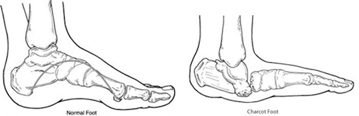
Eventually, the bone loss causes changes in the structure of the foot and areas of the foot collapse become severely deformed. When the collapse occurs to the midfoot, it rounds the bottom of the foot into a so called rocker-bottom foot deformity. Depending on the location of the bone break, the toes can start to curve under like claws or the ankle can become deformed and unstable. Sharp edges of bone may put pressure on the skin, creating the risk of chronic skin sores. The end result — a combination of bone disintegration and trauma – is Charcot foot.
Symptoms Of Charcot:
- A foot that is warm to the touch and noticeably warmer than the opposite foot
- Redness (may mimic infection)
- Swelling
- Pain or soreness
Some Charcot joints, such as the ankle, may heal with fibrous tissue and this may result in gross instability (“floppy foot”) that may predispose the person to foot ulcers and may be difficult to support with braces.
How Do We Diagnose Charcot?
Early diagnosis of Charcot foot is important to stop the foot from further destruction and collapse. X-rays and/or CAT scans are the mainstay imaging modalities to evaluate the bone destructive pattern. However MRI may cause confusion with osteomyelitis (bone infection) versus Charcot.
Treatment Of Charcot:
- Immobilization. Because the foot and ankle are so fragile during the early stage of Charcot, they must be protected so the weakened bones can repair themselves. Complete non-weight-bearing is necessary to keep the foot from further collapsing. The patient will not be able to walk on the affected foot until the surgeon determines it is safe to do so. During this period, the patient may be fitted with a cast, removable boot, or brace, and may be required to use crutches or a wheelchair. It may take the bones several months to heal, although it can take considerably longer in some patients.
- Custom shoes and bracing. Shoes with special inserts may be needed after the bones have healed to enable the patient to return to daily activities, as well as helping prevent recurrence of Charcot foot, development of ulcers, and possible amputation. In cases with significant deformity, bracing is also required.
- Activity modification. A modification in activity level may be needed to avoid repetitive trauma to both feet. A patient with Charcot in one foot is more likely to develop it in the other foot, so measures must be taken to protect both feet.
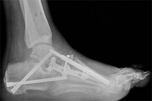
Surgical Intervention For Charcot:
In most case the deformity that is the result of a Charcot flare up will leave the foot in a poor position and predispose the patient for ulcerations, infections and amputations. Surgical options may include realignment osteotomies and fusion (correction of deformity), or ostectomy (removal of bony prominence that could cause an ulcer)